by Claude ROUSSEAU
ancien Président de la SFHAD
Ex-conservateur du Musée Pierre Fauchard
It’s with the Cnide School of Medicine that observation and concrete expression of facts take the place of incantations of magical medicine.

This concept blossoms with Hippocrate where clinical examination becomes an attempt to understand the patient before studying the disease and take everything into account: « What is possible to see, to touch, to hear, what is perceptible by sight, by touch, sense of hearing and smelling … »
The method of observation and the principles of the practitioner of the School of Cos largely contributed to the outstanding development of western dentistry.
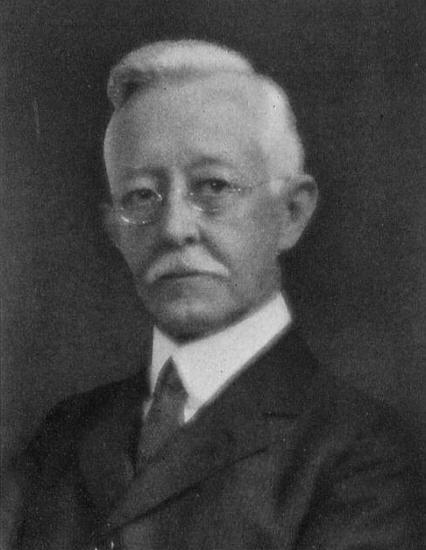
Wilhelm Conrad Röntgen
But with the announcement by Wilhelm Conrad Röntgen, who discovered in Januaary 1896 that the new radiation was able to cross opaque living tissues, we enter a new era of medico-dental diagnosis.
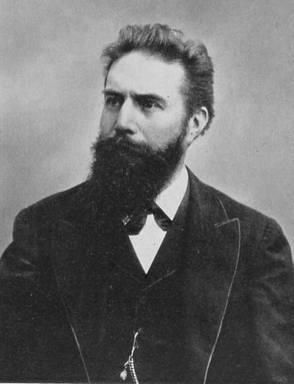
Born in Lennep on March 27, 1845, Röntgen works in Würzburg in 1870, in Strasbourg in 1872 where he becomes Privatdocent in physics in 1875.(1) (2)
In I879 he is professor of physics in Giessen in 1888 in Würzburg where he manages the Institute of Physics, dean of the university in 1893 and Nobel Prize in 1901.
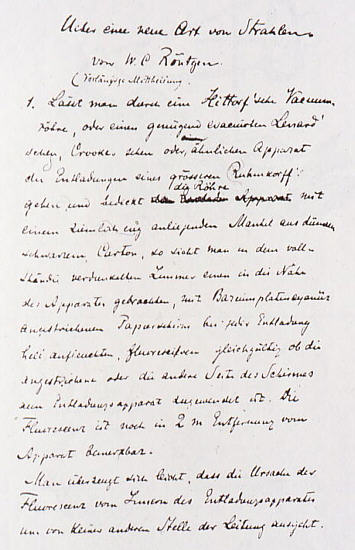
The genesis of his discovery is recorded in the seventeen paragraphs of a report entitled « Uber eine neue Art von Strahlen » (on a new kind of rays) he gives on December 28, 1895 to the secretariat of the Institute of physics and medicine of Würzburg.
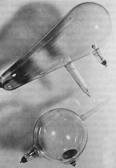
Decided to recapitulate Hertz and Lenard ’s works he gets several modified tubes of Hittorf and an induction coil outputting 20 amperes with a tension corresponding to 15 cm sparks, in his laboratory about May 1895.
In November 8, 1895, Röngen switch the light off. In the darkness of his laboratory, he notes that the screen saturated with barium platinocyanide, left at some distance away on a table, starts to emit light whatever the positions and the incidences of the sceen.
Seeking the cause of this phenomenon, he notes that the tube of Hittorf carefully wrapped in a sleeve of black paperboard he just happen to manipulate remained connected to the induction generator still under tension.
Röntgen has at once the intuition that his tube emits an unknown radiation able to cross the black paperboard and expose his screen by causing the phenomenon of fluorescence.
A first series of expériments shows him that this new ray calls X differs from the light. But, at the time of a new series of experimentations, he notes that X-rays are comparable with the light by
absence of deviation by the magnetic field, with the production of shadows and fluorescence and by the chemical effects resulting from the formation of a picture on a photographic plate even when this one is closed in the frame.
He proceeds then to a series of usual objets radiographs on photographic plates and finishes by radiographying the hand of his wife Bertha on December 22, 1895.
This historical X-ray appeared in the review « Nature » of London on January 23, 1896.
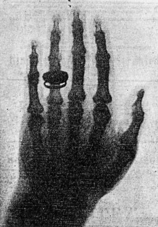
In January 23, Röntgen is invited by the physico-medical Institut of Würzburg where he shows the result of his research materializing by making X- ray of the hand of the famous anatomist Albert von Kolliker.
First applications of X-rays in Dentistry
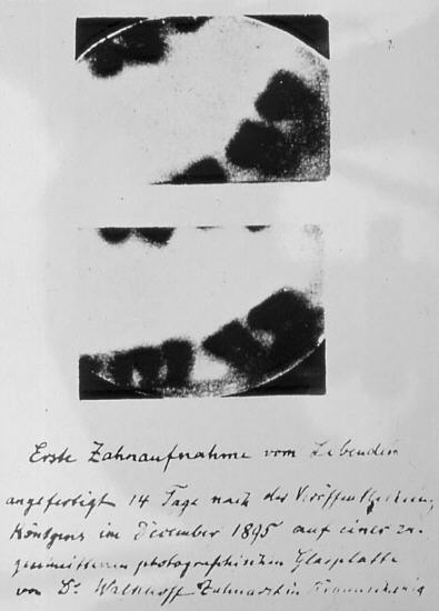
The forschungsinstitut für Geschichte der Zahnheilkunde shows in January 1896 two dental radiographs with the mention: » The dental X-rays of Dr Walkhoff, dentist in Braunschweig, made 14 days after the Röntgen publication in December1895 «
But if we refer to the publications of this practitioner (3) we learn that on the initiative of Dr.Walkhoff, Professor Giesel carried out on his friend these two intra-oral X-rays with the help of a small glass photographic plate wrapped in black paper and a sheet of rubber.
Walkhoff reports, that the exposure lasted 25 minutes and adds: « it was a true torture, but I felt a great joy at the sight of the results when I become aware of the importance of the Röngen rays for dentistry. »
On february 2, 1896 Professor König presents 14 radiographs at the Physical Society of Frankfort.
These 14 X-rays were published in March under the title: « 14 Photographien mit Röntgen-Strahlen von Prof W. König » and published by S.A. Bart, Leipzig in 1896.
The main point of this presentation refers to the exposure time reduced to 9 minutes. Schaeffer and Stuckert and more recently Streller describe the Professor König’s apparatus responsable for the important reduction in time exposure. (4) (5)
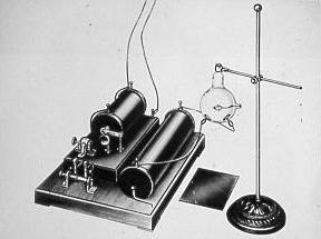
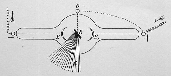
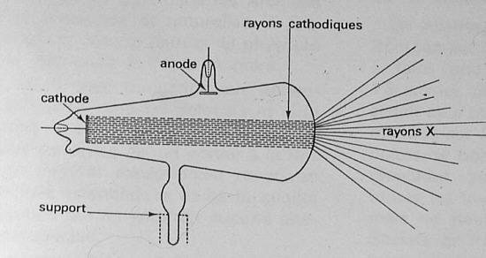
With the tubes of Crookes and Hittorf the cathodic beam spread on a straight line, perpendicular to the plan of the surface of the cathode, and runs into the opposite wall of the tube of glass. The results is a heating of the glass which softens the tube and makes difficult the maintenance of precise pressure.
The new tube of König differs from the precedent one by its anticathode which is made up with a platinum disc located at 45° of the of the convergence point of the cathodic beam. From this emergent point of the electrons, the X-rays spread out in every directions.
This device, called by the Anglo-Saxons « focus tube »or « reflecting tube » generate also a production of more penetrating X-rays and a noticeable improvement of the definition of the tissues upon the photographic emulsion.
Schaffer and Stuckert will bring back the duration of the exposure to 5 minutes by using the König’s tube.
During a meeting of the Central Association of Germans Dentists, Walkhoff, in his turn, shows some X-rays in april 1896.
In 1897, he exhibits for the members of the Academy of Sciences the earliest cranial X-rays made with the Professor König’s tube that underline a better definition of the film.(6)
The french experimentations
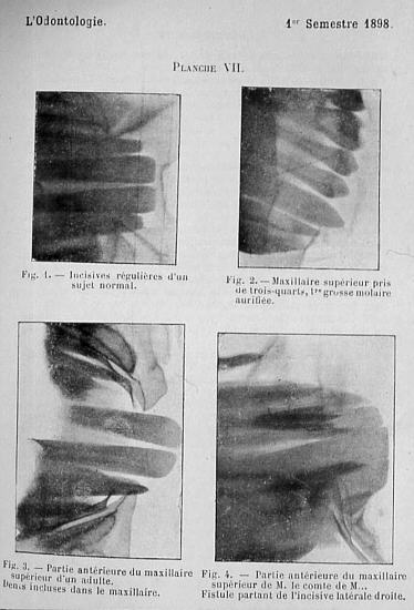
In France, the Professeur Béclère set up in 1897 the first laboratory of radiology in his department of the Tenon Hospital .
The first conference on X-rays are read by Godon and Contremoulins where they introduce their own technic and the practical applications with the Louis Richard Chauvin and Félix Allard’ X-rays. (7) (8)
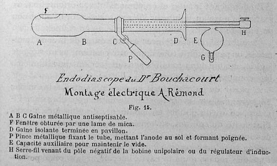
On february 7, 1899, le Dr Bouchacourt shows his « Endodiascope » drawn from the Dr Williams Rollins’ s « fluoroscope » (9)
The anglo-saxon’ s experimentation
If Frank Harrison (10) (11), in England, is considered the first user of the X-rays, in the United States, William James Morton, the son of the famous anesthetologist, publishes the first dental skiagraphs in the le Dental Cosmos of april 24, 1896.
They are claimed as the first X-rays published in the american litterature. (12) (13)
Dr. Edmund Kells is well-known for his important contribution to the popularization of electricity at the dental office.
But he is also regarded as the pioneer of the application of X-rays in the United States. His technic is developed in two important articles published in 1899.(14) (15)
The film, put in a small pocket wrapped with a double thickness of black paper and rubber, is set against the dental arcade perpendicular to the teeth to avoid any deformation.
For this purpose, he uses a film holder of his invention, maintained month closed to avoid any displacement and not, as you may believe, to avoid a radiodermatitis of the fingers of the operator.
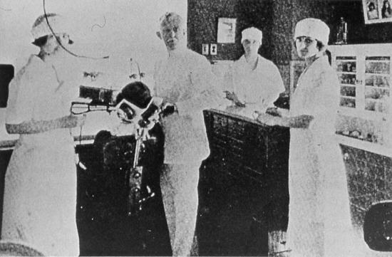
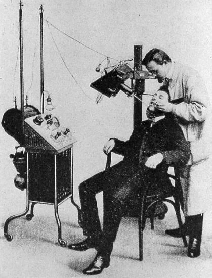
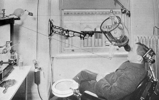
Dr. Stephens,who set up in Great Falls in Montana, describes the equipment of his office in 1897 in articles of the serie « Office and laboratory » published in the review « Items of interest ».(16)
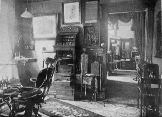
After an attentive examination of the picture of his office we discover a radiographic equipment, still very rare in 1897. Achieved in hand-made way, this lay-out includes :
-
a Crookes’s tube positioned horizontally under the wall mounted display and which is placed on a wooden tube holder, interdependent of a metal column fixed on a tripod.
-
a generator of X-rays, located at the extreme right of the photography, which includes an induction coil set on a table and a battery located on the the ground.
The dangerous high-tension wire which supply the Crooks’s tube are suspended from the ceiling and threaded in a ring in order to prevent a possible electrocution of the patient or the dentist !
In addition, there is neither voltmeter or amperemeter to measure the intensity of the current.
Thus, there is no device to control the quantity of X-rays produced.
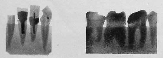
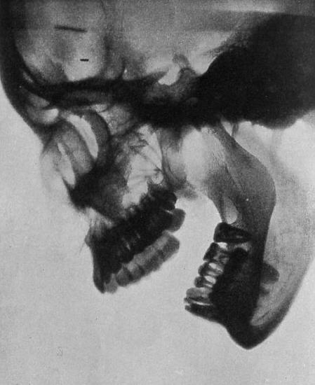
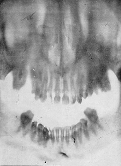
The two Dr Stephens’s X-rays.
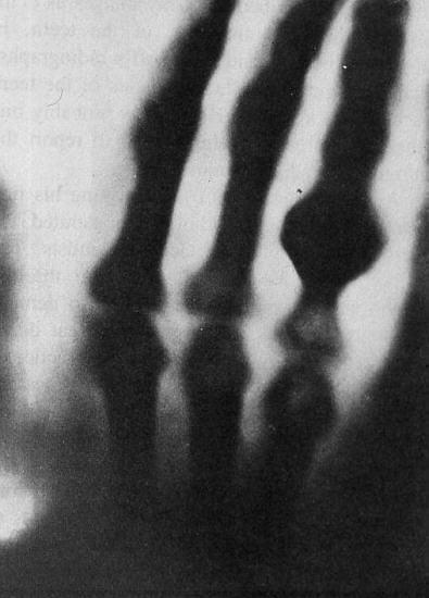
In spite of the archaïsm of this radiological apparatus , the two radiographs of the Dr. Stephens published in his article makes possible to see the broken broach passed throught the apex of the root (left) and a well defined exostosis of the root of the second molar (right)
Dr. Rollins was born on June 19, 1850 in Massachusetts. He gets the D.M.D degree from the Harvard dental school in 1875 when he was 21 years old, and the diploma of Doctor in medicine in 1879. (17)
As soon as the announcement of the Röntgen’s experiment , he begins an intensive work of research on equipments and the use of X-rays in Dentistry.
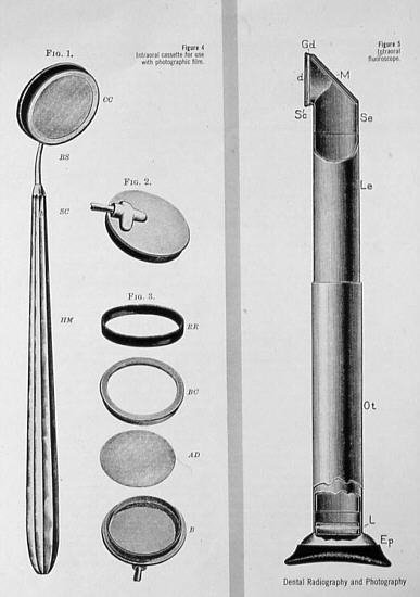
In July 1896, he invented an intraoral cassette and intraoral fluoroscope to look at the posterior teeth. However it is in the field of protection against radiation who he devotes from 1901 the majority of his researches.
In february 24, 1901, he publishes a resounding article entitled « X light kills ».(18)
In the consecutive accidents with the use of X-rays , Dr.Rollins affirms that the deletere agent relates primarily to the Röntgen radiations These publications are often accomodated with much scepticism.
Before his first publication of 1901 Dr. Rollins had already laid down the rules of protection against radiation.
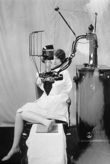
The first commercialy manufactured dental X-rays outfit
(Predecessors of Siemens)
The earliest manufactured dental X-rays apparatus.The X-rays tube was partially lead-shielded for protection but with exposed high-voltage wire. The output was 10 mA and 60 kV.
Dr Blum’s office of New-York
The Dr Blum uses a wall bracket X-rays tube holder with lead glass and protection shield.
The high- voltage wire which connects the generator to the tube are still not insulated. This type of equipment will mainly be used by american offices from 1905 to 1917.
This Ritter X-rays machine has still a dangerous exposed high-tension wire. This type of equipment was manufactured into the 1930 s.
Conclusion
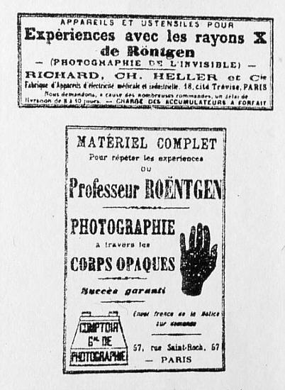
At this time everybody can buy a Crook’s tube with an induction coil at the « Comptoir general de la photographie
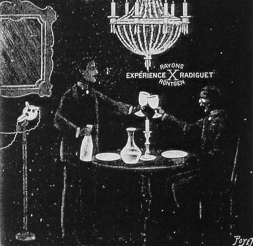
X-rays entertainment sessions are daily given at that time in private living rooms.
If the development of surgical and medical radiology were very fast, it was not the same in Dentistry.
Till 1900, a dozen of dental practitioners in the United States constitutes a small percentage
of dentists using this new technology.
This fact is due to the missing of specialised dental equipment and the missing of teaching which only starts in 1909 with Dr. Raper in Indianapolis. (19)
Price (20) announces that it is now established that the amounts of X-rays are cumulative.
Dr. Porter reported eleven cases of X-rays cancers, six of which proved fatal, as indicated by Dr Raper in 1907. (21)
In 1908 Lange (22) reports that X-rays operators have been made sterile from the influence of the X radiation..
Ketcham in 1911 (23) informs the profession of the death of the ingeneer Baur, first manufacturer of the « focus tube », victim of the repeated X-rays exposure .
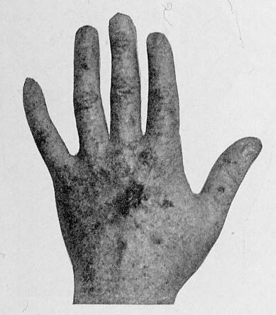
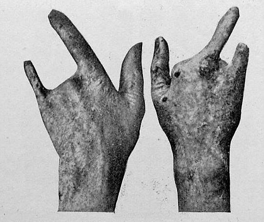
Regarding the Dr. Kells, he only become aware of this danger in 1912. (24) he reports, in its turn, many cases of amputations and even of death in the first users of the Röngen radiations.
Paradoxically, Kells thinks to be included in an other class of practitioners who would be comparatively immune against harmful effects of the Röntgen rays.
His opinion was unfortunately wrong, but after so much years of hazardous practice spent in his research laboratory, he will be, in his turn, reached by malignant lesions of the upper limbs.
Thus, after being amputeted of three fingers followed by the hand and the arm, it will put an end to his life on May 7, 1928. (25)
The name of Kells is consequently engraved on the list of the martyrs of radiological science.
Bibliography
1 Pizon, » La radiologie en France: 1896-1904 « , L’Expansion Scientifique Française, 1970.
2 Walkhoff O, » Altes und Neues vom Röntgenverfahren in der Zahnheilkunde « , Deutsche Monatsschrift für Zahnheilkunde, 1915, p. .8.
3 Walkhoff O, « Die erste Anwendung der Röntgenstrahlen und des Radiums in der Zahnheilkunde « , Correspondenz Blatt für Zahnärzte, Oktober 1928, n° 10, pp. 307-310.
4 Schaeffer und Stuckert, » Zahnaufnahmen mit Röntgen-Strahlen « , Deutsche Monatsschrift für Zahnheilkunde, Januar 1897, n°1, pp. 1-10.
5 Streller E, » Die erste Röntgenaufnahme von Zähnen « , Zahnärztliche Mitteilungen, 1965, n°19, pp. 947-949.
6 Walkhoff O, » Aufnahme der Gesichtsknochen mit Röntgenstrahlen « , Correspondenz Blatt für Zahnärzte, April 1928, n°2, pp. 97-99.
7 Godon et Contremoulins, « Les applications de la radiographie et de la radioscopie en art dentaire « , L’Odontologie, février 1898, VII, pp. 141-152.
8 Richard-Chauvin Louis et le Dr Allard Félix, « Application de la radiographie à l’Art dentaire « , L’Odontologie, février 1898, V-VI, pp. 152-155.
9 Dr Bouchacourt, » Introduction du tube de Crookes dans la cavité buccale « , L’Odontologie, avril 1899,V-VIII, pp. 311-318.
10 Harrison Frank, » The X-rays in the practice of dental surgery « , British Dental Journal 1896, p. 624-628.
11 Blackman Sydney, » Dental radiology – Past, Present, Future « , British Dental Journal, September 1959, p. 83-86.
12 Morton W.J., « Regular meeting of the New York odontological society « , The Dental Cosmos, May 1896, XXXVIII, n° V, pp. 401-402.
13 Morton W.J., » The X-rays and its applications in dentistry « , The Dental Cosmos, June 1896, XXXVIII, n° V, pp. 478-486.
14 Kells Edmond Ju., » Roentgen Rays « , The Dental Cosmos, October 1899, XLI, n° V, pp. 1014-1029.
15 Price, Weston, » The science of dental radiography (conférence au congrès international de Paris août 1900) « , The Dental Cosmos, May 1901, XLIII, n° V, pp. 483-503.
16 Stephens C.J.B., « Office of the Dr Stephens » Office and laboratory « , Items of Interest, 1897, XIX, n° V.
17 Dr Porter S. Sweet-William, Herbert Rollins, Dental radiography and photography, 1960, V, 33, n° 1, pp. 3-19.
18 Rollins William, » X-light kills « , Boston Medical and Surgical Journal, 1901, n°114, p. 173.
19 Raper H.R., » Notes on the early history of Radodontia – « , Oral surgery, 1953, n° 6, pp. 70-81.
20 Price Weston, » The technique necessary for making good dental – skiagraphs « , Items of Interest, 1904, XXVI, n° V, pp. 161-171.
21 Raper H.R., » The dangers of X ray « , Items of Interest, October 1912, pp. 725-730.
22 Lange Sidney, » The X-rays and their application to dentistry « , Items of Interest, 1908, XXX, n° V, pp. 522-535.
23 Ketcham A.H., » The radiography in orthodontic « , Items of Interest, 1911, XXXIII, n° V, pp. 281-302.
24 Kells Edmonds Ju., » Protection from the Roentgen Rays « , Items of Interest, Novembrer 1912, pp. 805-823.
25 Goaz P.W and White S.C., Oral radiology, The C. V. mosby C°, pp. 6-7.